Erectile dysfunction (ED) is a big issue in various populations worldwide. Erectile dysfunction is defined as a situation or inability to achieve and/or maintain penile erection that is sufficient for satisfactory sexual performance. It’s been observed that around 52% of the male population is affected with ED amongst which over 30% of young males are suffering from this condition. Scientific investigations have observed that the prevalence of ED is strongly associated with increasing age, making it a major health problem since with the increase of the aging population, the worldwide prevalence of ED has been predicted to reach 322 million by 2025.
Blood vessels play a prime role to keep erections firm. Under normal physiological circumstances, during sexual arousal nerves release chemicals that increase blood flow into the corpus cavernosum (erection chambers in the penis), the arteries in the penis expand to allow the inflow of blood which makes the erection firm. However, to keep the erection rigid, blood needs to stay in the penis until the man ejaculates. For this, penile veins constrict to trap the blood inside, which is known as veno-occlusion. At the end of sexual stimulation, the veins widen and blood flows back into the body. If the veins do not constrict enough, blood leaks back into the body, which results in softening of the erection, this phenomenon is known as a venous leak in which the erection gradually loses its firmness. Therefore, venous leak is a defect where a male is unable to have strong erections even with sufficient blood into the penis as a result of abnormal venous drainage in the penis corpora cavernosa, thus retarding the typical erectile capability of men. Venous leak is often listed under erectile dysfunction causes in younger men. Surveys have found that about 70% of the affected population by ED does not get treated therefore, men suffering from this condition experience shame and choose to be secluded in sexual relations. If you have concerns regarding being affected by this condition, contact your urologist, and get an early treatment since venous leakage is treatable. Although there are quite a few causes of erectile dysfunction, nonetheless, many non-invasive remedial options are available for such patients that will improve fertility and its progression.
Erectile Dysfunction Causes
There are many different underlying factors that can qualify as erectile dysfunction causes. Based on etiology, erectile dysfunction is divided into three major types: psychological (such as depression, stress, and anxiety), organic (related to vascular system, nervous system and endocrine system) and mixed.
When we talk about organic causes, defects in the physiology of vascular system are a common occurrence, in patients with venogenic erectile dysfunction, veno-occlusive dysfunction is recognized as underlying cause in most cases which can be due to either aging or injury related changes to the tunica albuginea, cavernosal smooth muscle dysfunction or excessive adrenergic input or shunts created during priapism episodes. There are several potential risk factors such as age, diabetes, obesity, cardiovascular disorders, hypertension, atherosclerosis, prostatectomy, pelvic radiation, Peyronie’s disease (development of scar tissue inside the penis), and androgen deprivation therapy that appear to be associated with organic ED.
Diagnosis and Treatment
In order to diagnose venous leak erectile dysfunction, your doctor will take your personal history, would likely do a physical examination, and recommend some tests such as Penile doppler ultrasound,certain male hormone and routine blood tests along with urine tests to rule out any doubts.
It is advisable for patients with venous leak erectile dysfunction to make lifestyle modifications and prevent physiological stresses to overrule these conditions and take adjunct medications. Furthermore, for patients suspected with ED are commonly prescribed phosphodiesterase type 5 inhibitors (PDE5Is) therapy which is not a curative treatment but rather symptom-relieving treatment and loses its effect in in a few months. There are surgical interventions also available in the market such as penile implant(recommended only in elderly and patients who had radical prostate surgery) and venous ligation surgery, which aims to repair the leaking veins(discouraged by surgical fraternity for recurrence and high failure rates).
Moreover, an interventional radiological approach that is endovascular treatment is a promising approach to treat patients with erectile dysfunction or venous leakage. Endovascular therapy with 3D precision-guided embolization of venous leak is a minimally invasive procedure and a safer alternative to surgical methods. For endovascular embolization, administration of local anesthesia followed by real-time fluoroscopic monitored puncture of a venous access is performed to reach the leaking veins and periprostatic venous plexus under sonographic and 3-D precision guided fluoroscopic guidance.
The non-invasive endovascular treatment can be performed by two different approaches, such as antegrade approach via deep dorsal penile vein and retrograde transfemoral venous approach via internal iliac veins. Although both approaches aim to embolize the periprostatic venous plexus along with other unusually shaped or abnormally functioning veins due to valvular incompetence, antegrade endovascular treatment is preferred since it provides a more direct route for catheterization of the periprostatic venous leaks.3D Precision guided fluoroscopic guidance helps the catheter reach the specific veins, following a diagnostic venogram embolization is performed using N-butyl-2-cyanoacrylate and ethiodized oil mixed in specific ratios depending on proximity, size and extent of venous leaks under continuous fluoroscopic control in neutral projection with optional oblique projection for precison guided embolization of incompetent pelvic veins.
This non-surgical treatment- 3D precision-guided embolization of venous leak is a complex and challenging procedure which requires high expertise in the said field. Its treatment in Pakistan was introduced by Dr. Imtiaz Ahmed in 2019, making him a pioneer. Dr Imtiaz Ahmad is an endovascular surgical specialist and an interventional radiologist who treats men suffering from venous leakage, erectile dysfunction and infertility issues that may have little hope left otherwise. His expertise in the field speaks volume for itself, which can be inferred by the accolades of his treated patients.
Uterine polyps, also commonly known as endometrial polyps are soft fleshy growths attached to the endometrium that is the inner wall of the uterus (or womb) and extend into the uterine cavity. Although uterine polyps have a low incidence rate compared to other reproductive ailments with every one in 10,000 females being affected. However, they can be absolutely worrisome when they straightforwardly affect fertility, pregnancy outcomes, and reoccur after polyp removal. These are somewhat similar to skin tags, which are normal tissues in essence but grow in an abnormal fashion and often flatten out to fit the cavity of the uterus. Endometrial polyps range from a few millimeters in diameter – such as a sesame seed – to several centimeters, like a golf ball or larger. They are attached to the uterine wall by either a large base or a thin stalk (or pedicle). Uterine polyps are commonly observed in 10-25% of women worldwide that are usually in their fifties. These are rare to find in women under 20 years of age or those who have surpassed menopause.
Polyps can occur anywhere in the body ranging from stomach, ear, and nose to different locations in the female reproductive system such as ovarian polyps. It is to be noted that there can be more than one polyp in the uterine cavity. Usually, polyps are contained within the uterus, however, occasionally, they slip down through the cervix (opening of the uterus) and into your vagina, blocking the opening. In such cases, they are referred to as cervical polyps and vaginal polyps, respectively. The vast majority of these polyps are usually noncancerous (benign), although in some cases they can be cancerous or eventually turn into cancer at the end.
Often times people misunderstand the difference between uterine polyps and uterine fibroids since both polyps and fibroids are similar in the essence that they grow on the uterine wall. Statistics show that about 80% of women experience at least one of the conditions in their lifetime. Uterine polyps are oval tissue growths that form in the lining of the uterus; whereas fibroids are benign growths that are found either outside or in the endometrial wall or within the uterine cavity (womb). Fibroids are likely to be developed in younger females, in contrast to polyps that are usually observed in middle-aged women.
Why Do Uterine Polyps Occur?
To date, the exact cause of endometrial polyps in women is not known. However, there are some theories that can best explain why they occur in the first place. These include:
Hormonal Imbalance: It’s been observed that polyps tend to grow when there is more of the hormone estrogen produced in the body.
Endometrial hyperplasia: It is the abnormal growth of the uterine lining. Since uterine polyps arise from the endometrial cells which grow during the menstrual cycle in preparation for embryo implantation. In case of absence of pregnancy, this lining sheds; however, due to numerous reasons, if the endometrial lining grows too much, it results in the formation of tissue outgrowth.
Genetic Factors: Various genetic factors have been found to be linked with the growth of uterine polyps, such as over expression of an enzyme found in the lining of the uterus called aromatase has been found to be one of its causative factors.
Risk Factors for Developing Uterine Polyps
The possible risk factors that increase the chances of developing uterine polyps are jotted down as follows:
Perimenopause or postmenopausal period
Hypertension
Obesity
Hormone replacement therapy or drug therapy
Uterine Polyps Symptoms
Uterine polyps are rarely symptomatic. However, in case of being symptomatic for these polyps, women show various signs and symptoms such as,
Irregular menstrual bleeding
Menorrhagia (heavy menstrual bleeding)
Bleeding after intercourse
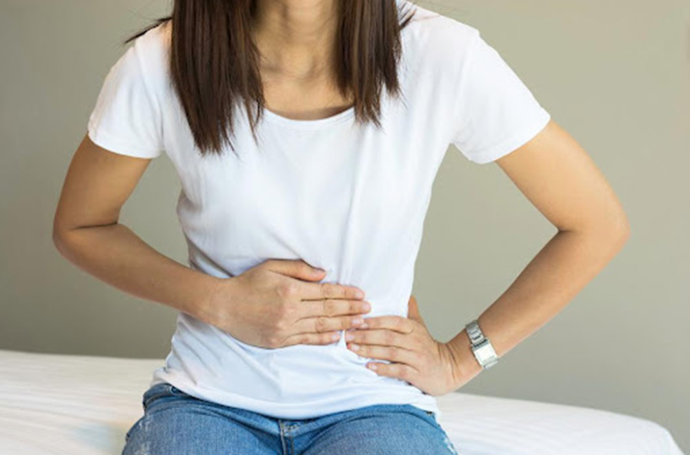
Cramping in the lower abdomen (dysmenorrhea)
Infertility or miscarriage
Uterine Polyps Diagnosis and Treatment
After your normal check-up and history, your medical specialist will likely recommend some tests to ensure if the condition correlates to uterine polyps or not. These tests may include vaginal ultrasound, hysteroscopy, endometrial biopsy, and curettage for confirmation.
If you are not symptomatic, then you shouldn’t worry too much about its treatment. However, polyps’ removal becomes a necessity if they are causing any aforementioned symptoms or if they are suspected to be precancerous or cancerous.
Medications: Certain hormonal medications such as drugs that help regulate the hormonal balance, like progestins or gonadotropin-releasing hormone agonists, could be used as a temporary treatment to relieve the symptoms. However, the symptoms will likely return after the medications are stopped.
Surgical removal: Polypectomy is a procedure to remove polyps. The most effective way to perform a polypectomy is through hysteroscopy, in which a small camera is placed inside the uterus using a thin, flexible, lighted telescope (hysteroscope) through your vagina and cervix for visualization of the uterine cavity. The procedure is performed under anesthesia to overcome any discomfort. During the procedure, it is possible to remove polyps from the uterus using a hysteroscope. Curettage is another surgical way to scrape uterine lining and removal of small polyps other than hysteroscopy. Other than that, hysterectomy is performed in cases uterine polyps are found to be cancerous, and thus the whole uterus is removed surgically.
Non-Invasive Solution to Uterine Polyps
Even though surgical removal is one of the available options for the affected females, however, it is not the common and most preferred choice since it involves either cutting you open or putting instruments into your uterus through the vagina. Moreover, the literature suggests that there are about 2.5% – 43.6% chances of recurrence after polypectomy. Additionally, in Asian societies and cultures, like that of Pakistan, fears regarding woman’s loss of ‘virginity’ are insurmountable. Therefore, preserving the virginity of an unmarried female is taken as the utmost priority so that it doesn’t affect their eligibility to get married or their future marital status and life. For this reason, many women remain either undiagnosed or untreated which ends up substantially affecting their quality of life or becomes a threat to their life, in general.
Here is the good news: there is a low-risk and non-invasive alternative procedure to surgical intervention known as 3-D Precision guided uterine artery embolization (UAE), which is now routinely available as an option. This is an outpatient procedure in which the uterine arteries that supply blood to the vascular polyps are located using real-time 3D fluoroscopic equipment. Blood supply to these polyps is blocked by injecting biocompatible particles into the bloodstream through a catheter at the wrist or groin, in which it leads to their ischemic infarction. As a result, they may be dislodged and excrete out from the uterus. This procedure has high success and minimal recurrence rate which eventually restores the quality of life of the affected individuals, taking them back to life.
The facility of 3-D Precision guided uterine artery embolization (UAE), is now available in Pakistan in its two major cities; Karachi and Lahore, by Prof. Dr. Imtiaz Ahmad and his team, who is a renowned interventional radiologist and endovascular surgical specialist with surgical training and extensive experience in Most Advanced Endovascular and Interventional Procedures.
Obesity is a complex disease which is characterized by abnormal and excessive accumulation of body fat. It is a condition that is defined as having a Body Mass Index (BMI) greater than or equals to 30 kg/m2. Over the past few decades, obesity has managed to become one of the major global public health epidemics that affects both children and adults. Therefore, it isn’t any more just a cosmetic concern, as it is accompanied by excessive health risks and medical problems that makes it a major cause of morbidity and a high rate of mortality worldwide. Along with its negative psychological manifestations it either causes, exacerbates, or adversely impacts numerous medical comorbidities and has now been recognized as a risk factor for several health ailments including diabetes mellitus, liver and cardiovascular diseases, musculoskeletal disorders such as degenerative joint disease and even malignancy. It is linked with several inherited and environmental factors, combined with lifestyle choices such as diet and exercise preferences. This explains why some people amongst us face difficulty in shedding the extra fat by varying degrees.
Obesity Treatments: What are your options?
There are several obesity treatment options in the market today which include lifestyle modifications, medical interventions, and surgical procedures. Where lifestyle modifications such as controlled diet and regular exercise can help in your journey to weight loss but only by 5% to 10% in case of either overweight or obese patients with low long-term efficacy, as reported by previously undertaken obesity research around the globe. To put more emphasis on this point, there are numerous studies that have shown that patients are likely to return to their original weight in about three years’ time frame.
Bariatric pharmacotherapy options are also available, but it requires you to fit a particular criterion to be eligible for using these medications. Currently, there are 6 FDA-approved anti-obesity drugs which mostly tinker with your nervous system, resulting in either reduced appetite or enhanced satiety. Unfortunately, the weight-loss efficacy of these medications is found to lie between only 3 to7%, and that too as adjuvants to other traditional therapies.
However, for patients that are either severely obese or those experiencing obesity-related comorbidities may require more aggressive and rigorous invasive medical management such as bariatric treatment or more simply put it, weight-loss surgery. However, it has been found to be associated with a relatively high morbidity rate in addition to the hefty costs that are required to undergo these surgical procedures. Yet, even after these cons, this approach towards weight loss has limited treatment outcome. There are several types of bariatric surgery options for you to undergo, but in essence, weight loss is achieved via reduction in the consumption of food or through either poor absorption of nutrients into the blood dream. Bariatric procedures when combined with lifestyle changes can however result in significant and sustained weight loss. Different types of bariatric procedures that are most commonly used include:
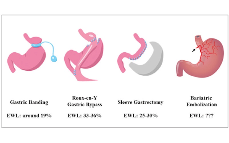
Gastric Banding – In this type of surgical intervention, a ring with an inner inflatable band around the top of your stomach is placed to create a small pouch. The gastric band makes you feel full much sooner after eating a small amount of food.
Sleeve Gastrectomy – In this procedure, the surgeon removes a major chunk of your stomach, leaving only a banana-shaped section behind therefore, this type of surgery cannot be reversed. The surgery limits the amount of food that can be consumed to a large extent that makes you feel full sooner.
Roux-en-Y surgery (Gastric Bypass) – This procedure allows your surgeon to staple the top part of the stomach which creates a small pouch, and then attach it to the middle part of the small intestine so as to reduce the intake as well as absorption of food from the gut.
Gammablock Bariatric Arterial Embolization
Luckily, there is an alternative treatment with lower morbidity rate and cost also available, termed as Gammablock Bariatric Arterial Embolization or simply Bariatric Embolization it is a novel technique first discovered and used by Dr Imtiaz Ahmad in 2017.It is a minimally invasive trans-catheter procedure that employs the endovascular approach. It aims to induce localized ischemia in the stomach to modulate the endocrine functions related to appetite that result in weight loss. Multiple clinical trials and studies have yielded exciting results that demonstrate appetite suppression and weight loss in patients after undergoing BAE. Clinical data obtained through research have shown no major or serious adverse events after the embolization procedure apart from nausea, vomiting and superficial stomach ulceration.
Gammablock bariatric arterial embolization is performed by Sr Imtiaz Ahmad under real-time imaging guidance. In this procedure, trans-arterial embolization of the gastric fundus via the gastric arteries i.e., the left gastric artery (LGA), gastroepiploic artery (GEA) and Short Gastric Arteries supplying the gastric fundus is performed using embolic microspheres to induce localized ischemia in the gastric fundus, which is aimed to decrease ghrelin (hunger hormone) production in the stomach, leading to appetite suppression and weight-loss.
If your weight loss journey has only seen the troughs, it is now time to culminate it with a non-invasive, high yielding treatment option near you. This facility of availing this embolization procedure is now available in Pakistan in its two major cities; Karachi and Lahore, using FDA-approved drug by Dr. Imtiaz Ahmad, who is a renowned interventional radiologist and endovascular surgical specialist. As a part of post-treatment practice, the patient can expect a follow-up up to 6 months to a year.
In this age, the concept that infertility is somehow associated with only a woman’s inability to conceive has become outmoded. It is now widely understood that 30-40% of infertility cases may be due to problems with male counterparts. This is owing to either ineffective sperm delivery as seen in retrograde ejaculation, altered testosterone levels, or abnormal sperm count that can be associated with a condition known as varicocele. Regretfully, there are cases which can’t be helped; however, on the brighter side, all hope is not gone with the availability of multiple infertility treatment options in the market. Perhaps, if you suspect being the victim of varicocele, it is best suited to seek immediate medical attention than to wait until the condition gets worse to the point that it can’t is untreatable.
Explaining Varicocele
The reasons associated with male infertility correspond to their unique reproductive anatomy. In men, the scrotum is a skin-covered sac that holds the testicles along with arteries and veins that deliver blood to the reproductive system. Varicocele is a clinical condition that causes the enlargement and dilatation of the scrotal veins. These veins form a vascular network known as pampiniform plexus in the spermatic cord, which ultimately connects to the vessels in the abdominal cavity and takes blood back to the heart. A varicocele is a varicose vein of the testicle and scrotum that is very similar to varicose veins that occur in the leg. It results in low sperm count and decreased quality, which is usually the main culprit behind infertility issues. It is deemed as the most common yet correctable cause of male infertility which has been associated with deleterious effects on spermatogenesis, sperm quality, testicular atrophy, reproductive hormones and pregnancy outcomes.
Varicocele raises the temperature of the testicles, which has mainly two effects. First, it affects the development and quality of sperm and second, it leads to a decrease in the production of testosterone, which is involved in the production of sperm. Moreover, the increased temperature is said to lead to a rise in reactive oxygen species (ROS), which damages the sperm DNA.
Symptoms Associated with Varicocele
It’s scary to be in oblivion, when you don’t know what is wrong with you while nothing is going your way. Following are the few symptoms associated with varicocele.
Pain: Where many patients do not find any scrotal discomfort. Some go through mild discomfort or severe pain when standing or sitting for prolonged periods due to the pressure that builds up in the affected veins, which is relieved when the affected person lies on its back.
Infertility Issues: Nearly 30% of couples that are facing fertility issues are diagnosed with presence of varicocele in the male counterpart. Infertility has been said to be associated with raised temperature in the scrotum, which affects sperm production.
Testicular Atrophy: Testicular shrinking is another giveaway of varicocele in the affected individuals.
Diagnosing Varicocele
Varicocele diagnosis is traditionally made upon physical examination, which is typically performed in the laying and standing position, wherein the scrotum is examined for visible swelling during rest and straining conditions. Along with physical examination, scrotal ultrasound, venography, and color doppler ultrasonography are also used for its diagnosis.
It is graded ranging from grade 0-3 depending on the clinical findings on palpitations and testicular size. Where grade 3 varicocele is easily identified through physical examination however, lower grade varicoceles are difficult to pinpoint, particularly in case of certain clinical comorbidities.
Varicocele Treatment
Below are listed some available options patients suffering from varicocele can opt;
Surgical Procedures: There are three types of surgical approaches that are typically used for its treatment, namely, laparoscopic surgery, open surgery and microsurgery. All of these options involve hospitalization, incisions, extended recovery time, scarring and recurrence potential.
Healthy Diet and Supplements: It’s been observed that foods that are rich in fiber and antioxidants are exceptionally good for varicocele patients in relieving the symptoms. Furthermore, supplements might prove to be beneficial as well since our daily diet lacks all the necessary nutrients therefore, taking these will allow the consumption of the nutrients that we are missing out on in appropriate amounts which will have their long-term effects.
Varicocele Embolization: It is a minimally invasive 3-D precision guided treatment first time introduced by Dr Imtiaz Ahmad in 2019 in Pakistan. Dr Imtiaz Ahmad is an American trained and highly qualified Endovascular Surgeon and interventional Radiologist.This procedure is performed using real-time fluorescent imaging to guide catheters inside the body through the Jugular vein and sometimes femoral vein. Using either mild IV sedation or local anesthesia, patients remain relaxed and experience minimal pain during the procedure.No surgical incisions are formed with no risk of scars. The blood flow to the affected vein is stopped by injecting FDA approved medications through the catheter, which embolizes the abnormal veins with defective valves and thus reduces the pain, pressure and high temperature in the groin and specifically along the testis.Through embolizing the vein, the blood flow is redirected to healthy vessels in the vicinity.
Varicocele Embolization has been first introduced as a 3-D precision guided endovascular treatment in two of the major cities of Pakistan; Karachi and Lahore. The embolization procedure is carried out using FDA approved medications by Dr. Imtiaz Ahmad, who is a world renowned interventional radiologist and endovascular surgical specialist. As a part of post-treatment practice, the patient can expect a follow-up for up to 6 months. There are numerous success stories of patients that underwent the procedure performed by Dr. Imtiaz Ahmed, whose lives are restored to a hassle-free state that they have been looking for a very long period of time and couples are able to have successful pregnancies and children.
Uterine artery embolization (UAE) is a great alternative to conservative surgical procedures like hysterectomy with its highly promising results, low recurrence, and considerably shorter hospitalization period.
Uterine artery embolization is a minimally invasive procedure that is performed to stop pelvic bleeding that might be caused by either uterine adenomyosis, uterine fibroids, cancerous tumors, trauma or injuries amongst other conditions. Among these, adenomyosis is a common pathophysiological condition in women of reproductive age that leads to distressing conditions which affect our daily performance and interactions. Although a major chunk of our population of approximately 20% to 80% are affected by this gynecological problem, adenomyosis still remains highly under-recognized and under-researched disease. Therefore, it is important to spread awareness regarding this condition and its treatment plans to help those that are suffering in silence.
Uterine Adenomyosis – Overview
Adenomyosis is an often-misunderstood benign gynecological condition in which the lining of the womb or uterus that should be on the inside invades and get embedded into the muscular wall of the uterus and is primarily seen in women of ages between 30 and 50 years.
Uterine Adenomyosis Symptoms
As all of us act differently in similar situations, women affected by adenomyosis may be completely asymptomatic, which allows the disease to go unnoticed until the case becomes severe. However, on the contrary, a bunch amongst us experience symptoms that vary in severity, making it hard for them to go on about their daily business without any discomfort and difficulty. Since the symptoms of adenomyosis overlap considerably with those of uterine fibroids, people tend to confuse one with each other. Adenomyosis symptoms that are most frequently noticed in the affected women include intense fatigue, menorrhagia (heavy menstrual bleeding), dysmenorrhea, pelvic pain, and irritation in the bladder due to inflammation caused by the shedding of the invasive adenomyosis tissues in the uterus. Other symptoms may include abdominal bloating due to uterus enlargement, dyspareunia (painful intercourse), anemia and iron deficiency due to heavy monthly bleeding, irregular menstruation cycle while some also experience fertility issues.
Uterine Adenomyosis Causes
Being as under-researched as it is contrary to the affected population, the definitive root cause and pathogenesis of adenomyosis is yet to be discovered. Regardless, researchers have pinned down quite a few theories that could be the culprit behind its origin.
Age: Since this condition has been observed commonly in women over 40 years of age, it has might be linked with the aging process.
Surgical Procedures: It’s been seen that women who have had children especially through caesarean delivery or have previously undergone any uterine fibroid surgery or any intrauterine procedure are more at risk of developing uterine adenomyosis.
Invasionof Uterine Cells: Adenomyosis could develop due to the invasion of endometrial cell into the uterine muscular walls due to either an incision made during uterine surgery or during normal physiological conditions.
Inflammation: Inflammation associated with tears and incisions made during normal childbirth.
Fetal origin of extra tissues in the uterine wall that set down before birth and grow during adulthood.
Metaplasia of stem cells in the uterus, which is the differentiation of one type of adult cell into another type.
Adenomyosis Treatment
Earlier to deciding the treatment regime, it is necessary that definitive diagnosis of adenomyosis is performed. This may include physical examination to check for any uterine enlargement and tenderness, along with different imaging techniques like ultrasound and MRI. There are two options for its treatment, medically and procedurally depending on different factors such as your symptoms, severity of the disease and personal preferences. Below are a few treatment plans described that are used frequently to treat adenomyosis symptoms.
Medications: Non-steroidal anti-inflammatory drugs (NSAIDs) are prescribed to relieve adenomyosis associated pain.
Hormonal Therapy: Hormonal therapies have been suggested to control symptoms such as heavy or pain during by prescribing medicines like levonorgestrel-releasing intrauterine device (a contraceptive method), aromatase inhibitors, Gonadotropin-releasing hormone analogs and estrogen-progestin therapy.
Endometrial Ablation: This is a minimally invasive procedure in which the lining of the uterus is destroyed.
Hysterectomy: If you have surpassed child-bearing age or are ready to undergo surgery that removes the whole uterus to relieve the symptoms once and for all, then hysterectomy is another option.
Uterine Artery Embolization: Also known as uterine fibroid embolization (UFE), is an innovative outpatient procedure that has been established in 1995 as a beneficial treatment option for symptomatic uterine fibroids. Since then, the procedure has been established and is also used for the treatment of adenomyosis through adenomyosis embolization, which has shown favorable outcome.
It is a highly effective and essentially pain-free procedure performed by an interventional radiologist. Local anesthesia at the groin is applied, which may not even sting as prior to giving local anesthesia anesthetic spray is used over the access site.Specialized micron size particles are injected through a tiny tube inserted into the uterine artery that block the blood flow to the adenomyosis affected area. The procedure is guided through real-time fluoroscopy to direct the tube. The micron size particles then aid in shrinking down the invasive muscular growth that is the characteristic of adenomyosis by cutting off the blood supply to the affected area. Soon after embolization, pain (menstrual pain) may be experienced, however it will subside. You could feel the symptoms alleviate after the procedure. Being minimally invasive in nature, risks associated with surgical procedures such as wound infections and post-procedure complications are eliminated.
Uterine Artery Embolization is now available in Pakistan for the first time since 2017, as a 3-D precision guided endovascular treatment in two of its major cities; Karachi and Lahore. The embolization procedure is carried out using an FDA approved devices and medications by Dr. Imtiaz Ahmad, who is a renowned interventional radiologist and endovascular surgical specialist. As a part of post-treatment practice, the patient can expect a follow-up up to 6 months. There are numerous success stories of patients that underwent UAE procedures performed by Dr Imtiaz Ahmed that speak for his expertise in this area without compromising your fertility.