Spider veins are a medical condition affecting adults (women in particular). Numerous pain management specialists treat patients with spider veins to reduce the risk of having varicose veins. At the same time, they strive to enhance the visual appearance as well.
Spider veins are medically referred to as spider telangiectasias. It is a chronic dilation of the capillaries and other small blood vessels. They are usually smaller than varicose veins.
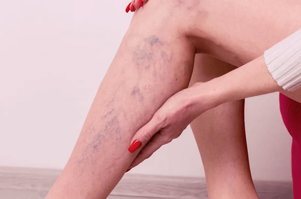
Spider veins are visible through the skin. They can create severe health conditions, such as skin ulcers, phlebitis, and blood clots. Let us read more to examine spider veins causes and how to treat the condition.
Understanding Spider Veins
This issue occurs from the enlargement of a group of small blood vessels visible through the skin’s surface. These veins usually appear as a red, blue, or purple sunburst (thus the term spider web).
Spider veins and varicose veins often co-occur. They are frequently visible on the face and as broken capillaries on legs. But they usually lie beneath the surface of the skin. However, they can appear anywhere on the body. They are also common in people above the age of 50.
Though they do not pose a health hazard, they can, however, produce a dull aching phenomenon in the legs due to prolonged standing. They can also be an indication of a more severe venous ailment. The incidence of spider veins increases with age, affecting slightly more women than men.
The modus operandi of Spider Vein Biology
The human body’s vascular system forms an intricate network of blood vessels that serves as the foundation for the development of spider veins. Understanding this process helps many people understand why they develop and how it affects their circulation.
Function and structure of the human vascular system
Blood flows through the human circulatory system in two ways: first, the arteries carry it away from the heart, whereas the veins return it. The veins contain specialised one-way valves that guide blood towards the heart. It prevents the blood from flowing backwards.
These valves work in conjunction with other muscular pumping mechanisms to ensure that blood continues to flow correctly. It is required when the legs need to move against gravity.
Formation of Spider Veins at the Cellular Level
Spider veins (spider telangiectasias) begin with abnormalities in the horizontal vascular plexus of capillary loops present in the skin. These damaged vessels can start from either arterial or venous origins.
Venous spider veins appear raised and have a bluish-purple color. They measure 1 to 3 millimeters in horizontal length. In contrast, arterial spider veins appear flat and pinkish-red in color. Their diameter ranges from 0.1 to 1 mm. The formation process involves numerous key mechanisms:
- Endothelial inflammation and vascular neogenesis occur because of local anoxia.
- Weakened vein walls due to an increase in blood pressure.
- Disruption of standard blood flow patterns.
The causes of spider veins
Spider veins can occur due to factors such as aging, sun damage, heredity, pregnancy (which can cause spider veins), hormonal influences, and trauma, all of which are known to be contributing factors. The exact cause is yet to be determined. Spider veins during pregnancy are visible due to hormonal changes. They can vanish after that or with the help of medications.
Spider veins are the outcome of structural abnormalities in the blood vessels. Veins take blood back to the heart from other areas of the human body. They use a series of one-way valves to avert the backflow of blood.
For numerous reasons, these valves can become defective. When the valves in the veins do not function correctly, blood backs up, leading to increased blood pressure. This pressure weakens the walls of blood vessels and hence the veins enlarge (dilate).
Numerous factors induce spider veins in a person, especially:
- Aging.
- Patients with a history of blood clots.
- Conditions (tumors, constipation, tight clothing) that cause pressure in the abdomen.
- Exposure to the sun and ultraviolet rays (visible in light-skinned people).
- Heredity.
- Obesity.
- Hormonal influences during puberty, pregnancy, and menopause.
- Postmenopausal hormonal replacement.
- Usage of birth control medications and pills.
- Occupations involving a lot of standing (nursing, hairdressing, teaching, and industrial occupations).
- Spider veins occur during pregnancy. But after childbirth, those veins gradually fade.
- Any past vein surgeries can also cause the condition.
- Injury or trauma to the skin.
Symptoms of Spider Veins
Spider veins typically have no noticeable signs or symptoms, except for a worrisome cosmetic appearance. But they can, at times, trigger restless legs, swelling, and throbbing. In some instances, they can become painful and can cause blood clots or skin sores. These symptoms often worsen due to prolonged sitting or standing. Affected individuals may also develop issues such as skin discoloration and skin ulcers.
Some people have asked the question, “Why are my veins so visible on my hands?” One of the key reasons is that spider veins have a web-like appearance on the skin. They can be red, blue, or purple. Here are other symptoms.
- An uncomfortable feeling in the legs.
- A Rash.
- Swelling.
- Cramps, aching, or throbbing.
- Restless legs.
- Itching is taking place around the veins.
Spider veins rarely cause any complications. However, some individuals affected by this condition can develop skin ulcers. These open wounds are often found on the lower leg and can lead to infections of soft tissues. Consult a physician for medical attention if:
- The veins are warm to the touch and quite tender.
- The veins cause pain upon touch.
- Development of rashes, sores, or ulcers on the skin.
- The spider veins bleed.
Diagnosis of Spider Veins
A qualified medical professional, such as a physician, can diagnose spider veins through a careful examination of the affected areas. For instance, physicians may want to examine the legs when patients are seated and also when they are standing.
The exam will consist of a visual inspection and palpation (i.e., touching) of the affected areas. Attention will be given to regions having redness, swelling, skin discoloration, and skin ulcers. Additionally, physicians would like to know the medical history of patients, especially the following:
- Recent illnesses or underlying medical conditions (like a heart condition or history of blood clots).
- Any medications or supplements the patient is taking (aspirin, blood thinners, herbal supplements, nonsteroidal anti-inflammatory drugs (NSAIDs)).
- Allergies.
- Previous treatments for spider veins and their outcomes.
In addition to a physical examination and a comprehensive medical history, physicians may order diagnostic tests to assess blood flow or detect blood clots. They can include the following:
- Duplex Ultrasound: A vascular ultrasound to check blood flow and vein structure (it uses two kinds of ultrasound).
- Color-Flow Imaging: Also known as triplex ultrasound, it is similar to the previous one. However, it utilizes color to depict the blood’s flow.
- Venogram: This is an X-ray test that involves the injection of dye into a vein to create an image of how blood flows through the veins.
- Magnetic Resonance Venography (MRV): This diagnostic procedure utilizes a combination of large magnets and a computer to examine the veins. Again, it injects a dye into the veins to see them better.
Treatment for Spider Veins
Spider Veins usually do not require treatment. However, many patients seek treatment for cosmetic reasons. If left untreated, they can develop into varicose veins. Moreover, not every person with spider veins experiences pain or other symptoms.
There is no proven way to reduce the appearance of spider veins. Various home treatments can reduce the appearance of spider veins. Conservative measures help prevent potential complications.
We will examine both medical and non-medical treatments for spider veins.