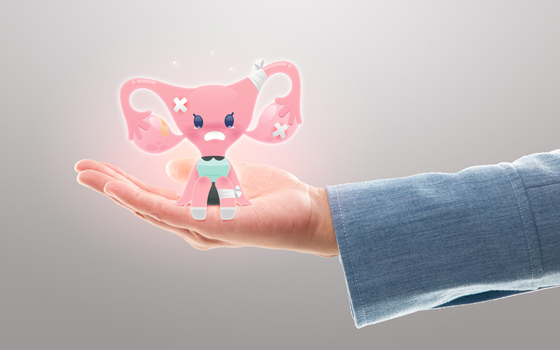
Uterine fibroids may directly or indirectly affect your married life, especially if you want to start a family. Get well versed with the treatment for fibroids
Uterine Fibroids and its consequences
Marriage is a social institution that officiates a relationship between a man and a woman. One of the greatest joys in life comes from the birth of a child. Becoming a parent changes a person on a whole and is an experience that matches none other. Surely, after marriage, almost every couple looks forward to appreciating this blessing. Unfortunately, issues regarding infertility have become more common now as people continue making unhealthy lifestyle choices. It is owing to advancement in science, that the causes are now better understood, and treatments/alternatives are also available. One of the conditions that may lead to female infertility is of uterine fibroids, however, successful pregnancy with fibroids is also not uncommon.
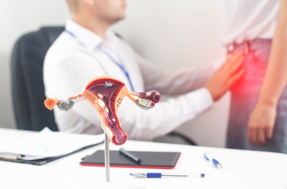
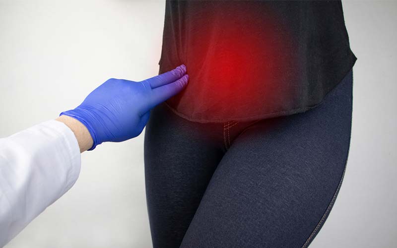
Uterine fibroids are one of the most common forms of tumors that develop in the reproductive system of women. These muscular growths are almost always benign and pose no serious threat. Fibroids in women are common in all age groups once girls reach puberty. Often, these benign masses go unnoticed owing to their asymptomatic nature. The asymptomatic uterine fibroids are of lesser concern than the ones that pose severe symptoms. Any aberration from characteristics of a normal menstrual cycle should alert a woman instantly. The signs and symptoms of uterine fibroids include heavy menstrual bleeding and extremely painful periods.
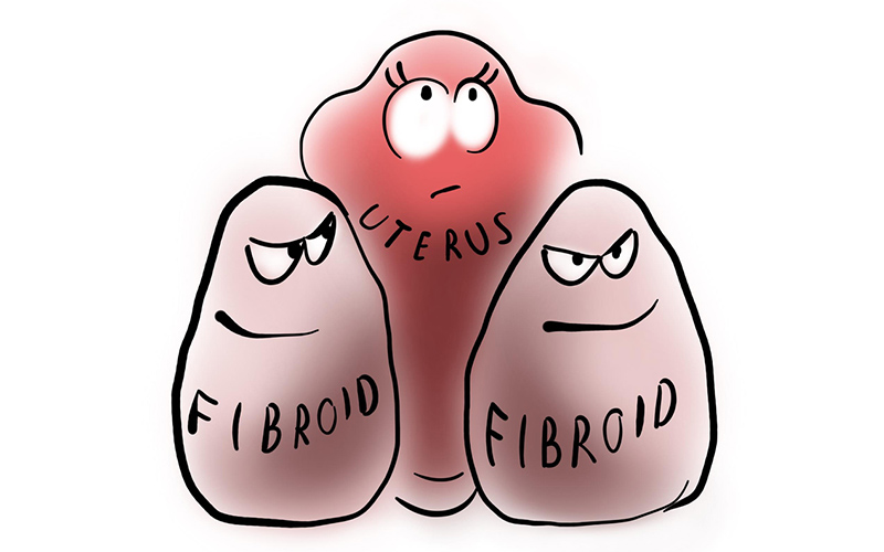
Adenomyosis is also another disease of the female reproductive system wherein a suffering individual faces heavy menstrual flow and painful periods. Since fibroids and adenomyosis have similar symptoms, adenomyosis is often wrongly diagnosed as fibroids. Fibroids vary in size and number and the symptoms are proportional to these variations. For example, multiple uterine fibroids are often so problematic that they may expand the uterus to a point that it touches the rib cage. Similarly, fibroids that are larger in size will cause more trouble, severe symptoms and will need immediate medical intervention.
The cause of fibroid in women is not known but it is certain that a few factors increase the risk of developing fibroid. These include maintaining an unhealthy lifestyle, hormonal imbalance, and being genetically predisposed to developing uterine fibroids. It is studied that uterine fibroids are more common in women over the age of 30 and teenager fibroid patients or fibroids in adolescents is a rare occurrence.
Treatment Options: Old vs New
Uterine fibroids that are symptomatic and larger than 10mm in size, may be treated surgically. In traditional gynecology, uterine fibroids causing complications are treated by hysterectomy, which is the removal of uterus. On the other hand, myomectomy may also be performed, wherein surgical intervention removes only the fibroids in the uterus.
Fibroid in unmarried girl or women who want to conceive may not want to go for uterus removal/hysterectomy. That said, myomectomy also adds to the misery with its high recurrence. Therefore, the best treatment for fibroids with high success rate is the non-surgical uterine artery embolization method, which may also be better known as 3-D precision guided treatment.
Dr Imtiaz Ahmad, an endovascular surgical specialist and interventional radiologist, is the pioneer of this treatment and has made this treatment available in Pakistan for the very first time since November of 2017.
3-D Precision Guided Endovascular Treatment
The 3-D precision guided treatment is an umbrella term that encompasses a handful of procedures, out of which uterine artery embolization alone, constitutes 60-70%. The entire procedure uses real time 3-Dimensional Fluoroscopy which is a type of X-ray that shows live imaging in 3- Dimensions. Live imaging helps in precisely locating the fibroids. The procedure begins with administering a local anesthesia in the form of “Cold Spray” applied to the skin of the groin area to numb it. Conscious sedation is achieved via IV in which the patient remains awake and relaxed. Avoiding any skin incisions or cuts, micro catheter is introduced through the femoral artery in the leg. The catheter is guided towards the uterine artery through which the blood to the uterus is supplied. Serial 3-Dimensional images are obtained followed by placement of specialized/engineered micro-catheters, if needed to complete the embolization.
In this way, the embolization procedure causes the blockage of blood flow to the fibroid(s) or adenomyosis. Additionally, Embolic agents are small micron sized particles that are passed through these catheters, these particles are engineered /programmed to block blood supply only to fibroids or adenomyosis while sparing the normal uterine walls. Another major component of the procedure comprises intra-arterial pain management done by intra-arterial infusion of carefully titrated medications directly into the fibroids/adenomyosis with super selective catheters selectively placed within the uterine fibroids or adenomyosis.
The patient is ensured more comfort via fluoroscopic guided nerve block which is effective for at least 18-20 hours post procedure. Post embolization syndrome which is an expected sequelae after any embolization procedure is dealt with there and then by infusion of specific medications directly into the fibroids/adenomyosis. All in all, the procedure takes around 45-60 minutes and has a high success rate amongst patients of all age groups. It is often difficult to relocate the access site since no incisions are made during the procedure. The patients are usually discharged only a few hours after the treatment in outpatient settings or are kept overnight under observation with a 23-hour admission. As part of post-procedural practice, the patient can expect a follow-up for a few months, especially if the procedure was also aimed at reversing infertility.